Prof. Maximilian Jösch
Neuroethology – Life Sciences, Institute of Science and Technology (IST), Austria
Background
Prof. Jösch is the head principal investigator of a functional imaging neuroscience group who are currently studying retinal processing in mice with a novel imaging method. The group wants to understand how the brain receives information about the surrounding world and how the information is processed and computed by the brain, using the retina as a window through which to study the brain. Many neurological diseases can be spotted early in the retina before they occur in the brain, including diseases like Parkinson’s and autism, which allows these diseases to be studied in a very controlled fashion.
They use a custom-built microscope imaging system to interrogate the function of live mouse retinal tissue via calcium (and potentially voltage) imaging.

Challenge
This kind of functional calcium imaging requires very high-speed imaging, with Prof. Jösch wanting to sample across the entire sensor at 100 fps in order to observe the fast dynamic functional processes in the retinal tissue.
As Prof. Jösch states: “We need to be sensitive and we need to be fast, if we are not sensitive enough we would need to have more light, and with more light we are basically destroying the tissue… since we are doing live imaging we want to avoid cooking the sample through direct light exposure.”
As the imaging takes place across live retinal tissue the largest field of view possible is desired but Prof. Jösch and team are more interested in function than anatomy: “Our main concerns are the signal to noise of the functional response so we can extract the information we require.”
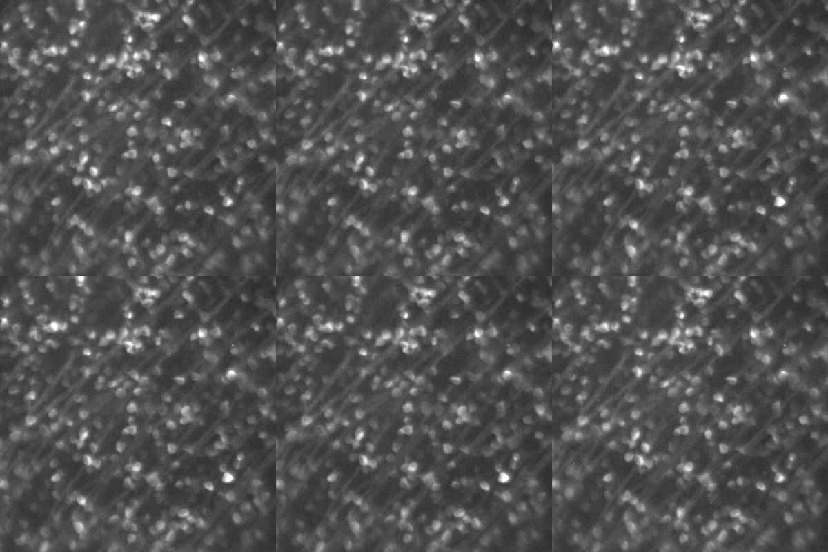
We wanted something that was more sensitive and faster, which is why we changed to [the Prime 95B], and the data looks pretty nice.
Solution
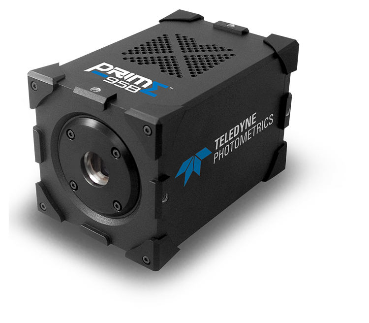
With the speed, sensitivity, and field of view of the Prime 95B, it makes a good solution for calcium imaging and functional neuroscience. As Prof. Jösch mentions: “We wanted something that was more sensitive and faster, which is why we changed to this system [Prime 95B], and the data looks pretty nice.”

