Dr. Guilherme Testa-Silva
Institute of Molecular and Clinical Ophthalmology Basel (IOB), Switzerland
Background
Dr. Guilherme Testa-Silva is head of Physiological Technologies at 1OB, part of a group committed to developing groundbreaking therapies for restoring sight.
Dr. Testa-Silva told us more, “We draw from areas such as molecular biology, clinical science, and physiological technologies. Our work includes understanding how the eye and the visual system function at a molecular and cellular level, and how alterations in these systems lead to vision loss. The ultimate aim is to develop new therapies that can restore sight, improve the quality of life for those with vision impairments, and contribute to the overall advancement of ophthalmic science.”
“To support our work, we utilize a range of state-of-the-art imaging technologies, including cameras for imaging retina structures; adaptive optics imaging; and two-photon microscopy for observing cellular and tissue properties in the eye. Furthermore, we continue to explore the application of genetically encoded voltage sensors, which allow us to study the electrical activity of excitable cells, giving us greater insights into the mechanisms of sight at a microscopic level.”

Challenge
“Low-light levels also pose a challenge. Given that certain procedures must be conducted under minimal light conditions to prevent phototoxicity or because the photoreceptor cells in the retina are sensitive to light, our cameras need to be highly sensitive to capture quality images in these conditions.”
“Lastly, there’s the challenge of imaging depth. To fully understand the complex, multi-layered structures of the eye, we require cameras capable of capturing images at varying depths, which can be quite a technological hurdle.”
“These challenges can significantly affect our imaging work. The inability to capture large fields of view might limit our understanding of the overall retinal structure and cellular interactions. Similarly, cameras with inadequate speed could miss rapid physiological events, reducing the accuracy of our data. Subpar sensitivity in low-light conditions might compromise image quality, while limitations in imaging depth could restrict our knowledge of deeper retinal structures.”
The Kinetix is an invaluable tool, it greatly enhanced our field of view, we can capture the retina in a single frame … the high speed has been crucial in capturing rapid physiological events.
Solution
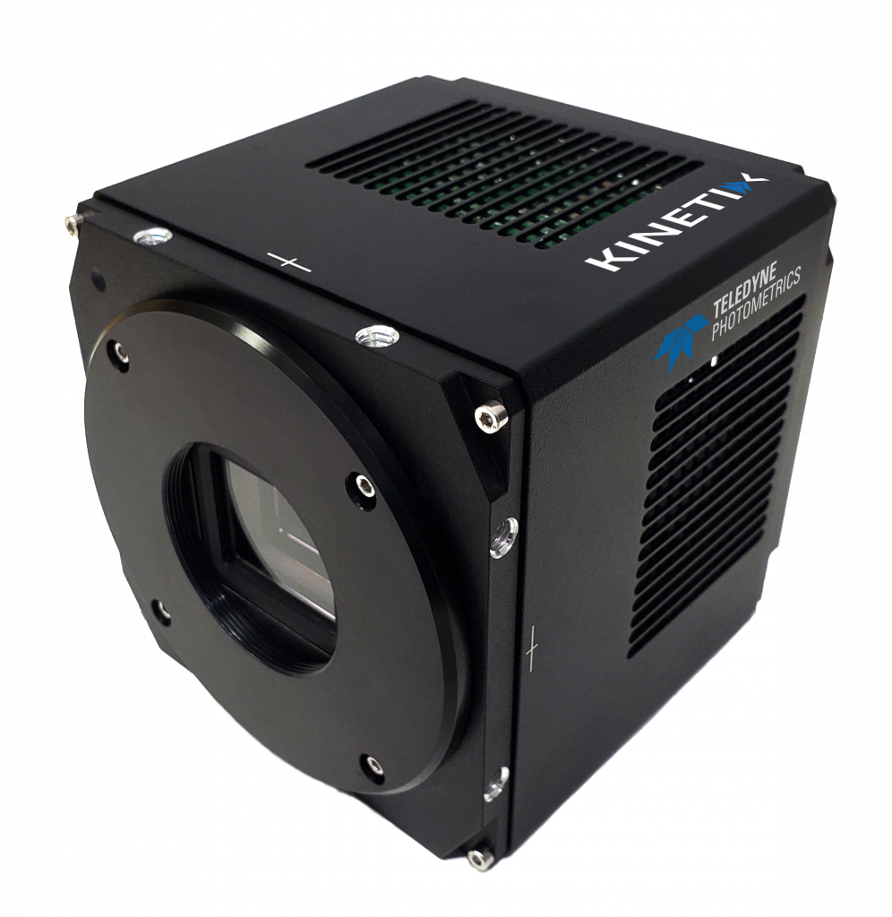
The Kinetix sCMOS is an ideal solution for this application, answering the need for large field-of-view imaging at high speeds in low-light conditions. Imaging at 500 fps across the entire 10 megapixel, 29 mm sensor, the Kinetix is well- suited to capturing events across large, dynamic samples.
Dr. Testa-Silva described his experience with the Kinetix, “The Kinetix camera has been an invaluable tool in addressing some of our primary imaging challenges. With its larger sensor size, it has greatly enhanced our field of view, allowing us to capture a comprehensive image of the retina in a single frame. Its high pixel resolution has also helped us obtain detailed images of individual cells and intricate structures.”
“Our experience with the Kinetix has been largely positive. The camera’s high speed has been crucial in capturing rapid physiological events. Its high quantum efficiency has proved advantageous in low-light conditions, enabling us to acquire high-quality images even in minimal light.”

