
Live Cell Imaging
Live cell imaging gives us the ability to observe dynamic processes in cells, tissues, or whole organisms as they happen. Compared to fixed cells, live cells provide us with information about the changes that occur in the cell during processes necessary for life. This includes everything from cell division to cell migration, movements and transformations of organelles and calcium imaging.
Our range of cameras for live cell imaging ensures that you’ll find the combination of specifications that match your requirements.

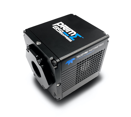
Prime BSI Express
High sensitivity, 95% quantum efficient, sCMOS camera with 6.5 µm pixels and 1.0 e– read noise.
The back-illuminated Prime BSI Express is particularly suitable for low light fluorescence imaging. The high quantum efficiency combined with the balanced 6.5 µm pixel size allows for Nyquist sampling with the most popular objective magnifications used for live cell imaging.
With live imaging possible at 95 fps full frame, you can rapidly find and focus on your sample and capture images using the shortest exposure times to minimize sample photodamage.


Kinetix
High sensitivity, 95% quantum efficient sCMOS camera with an incredibly high 400 fps full-frame speed and a massive 29.4 mm diagonal field of view.
When speed is needed, the Kinetix significantly outperforms typical sCMOS devices. With a full-frame speed of 400 fps, it’s possible to image live cells with greater time resolution than ever before.
The huge 29.4 mm square sensor of the Kinetix is designed to increase throughput, maximize the amount of data captured in a single frame and take full advantage of new, larger field of view microscopes.

Iris 9
High resolution, 4.25 µm pixel sCMOS camera with 9 million pixels.
Maximize resolution with the Iris 9, perfect for when the highest level of image detail is required. Live cell imaging is possible at over 30 fps full frame with a high, broadband quantum efficiency to image dyes with the most challenging emission wavelengths.

Customer Stories
Organoid Light Sheet
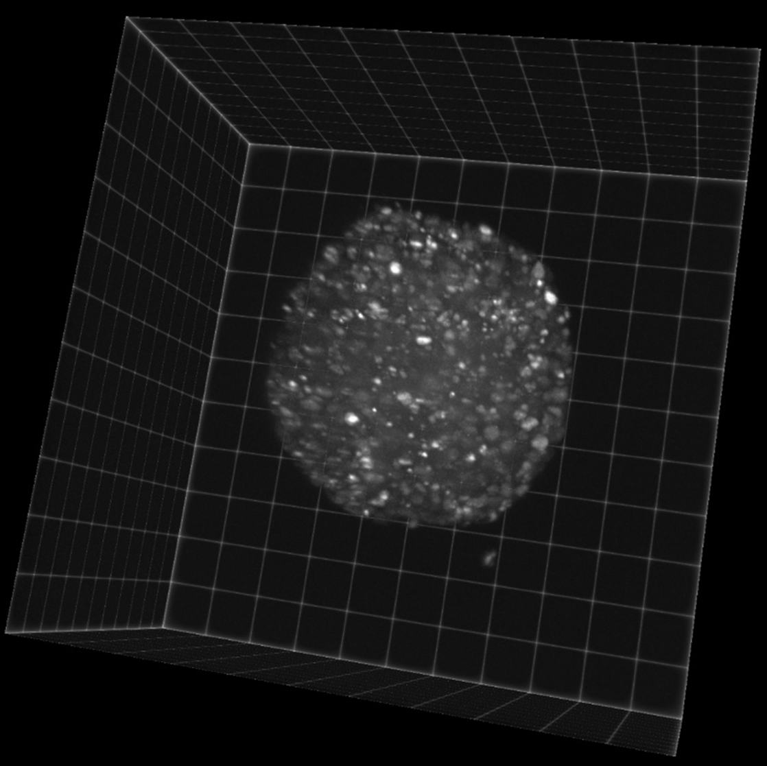
“So far every sample I have imaged fits into one field of view, and [the Iris 15] works great, it was easy to setup and image with.”
Single Objective Light Sheet
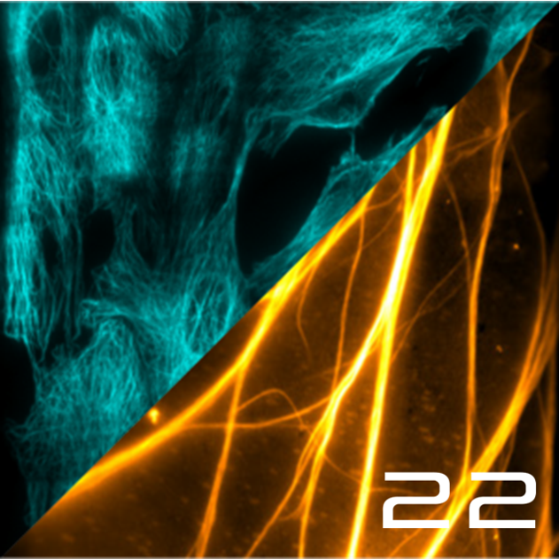
“”I really like the Kinetix22, it’s the best camera I have ever used on a system!””
High-Speed Calcium Imaging
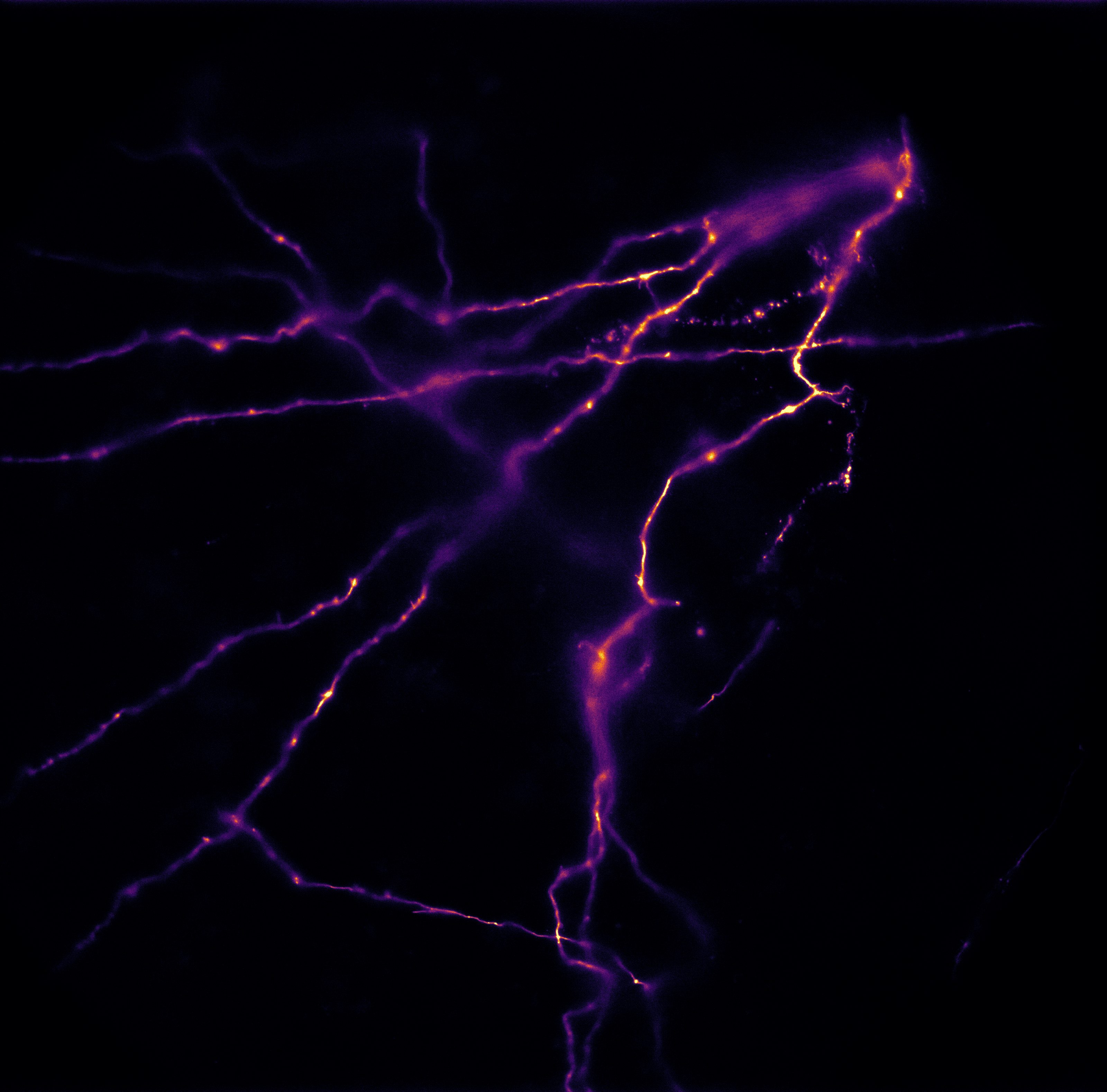
“The low noise and high readout speed of the Kinetix allow us to capture more photons while imaging at a faster rate.”