Prof. Dirk Geyer, Martin Richter (M.Sc.), Adrian Breicher (M.Eng.)
Laboratory for Optical Diagnostics and Renewable Energy (ODEE), Department of Mechanical and Plastics Engineering, Darmstadt University of Applied Sciences, Darmstadt, Germany
Background
The group of Prof. Dirk Geyer, including Ph.D. students Martin Richter and Adrian Breicher, work towards the decarbonization of energy conversion. They told us about their research, “Our main research field is the combustion of promising new fuels for the future such as hydrogen and ammonia, which, unlike methane, contain no carbon in their molecular structure and therefore produce no CO2 emissions in the combustion process.”
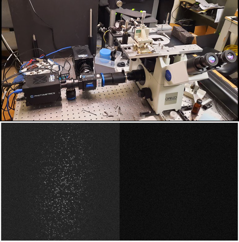
“In our recent work, we are looking at the co-firing of methane (CH4), the main component of natural gas, and hydrogen (H2). We have many combustion systems operating with natural gas, and we could substitute some of the CH4 for H2, but this has effects on combustion. To understand the fundamentals of these effects, we investigated laminar flames with reduced complexity, such as Bunsen flames: if we burn pure CH4 we observe a smooth flame cone, if we add certain amounts of H2 cellular structures start to appear, so we are looking into the structure of these with planar laser-induced fluorescence (PLIF).”
“PLIF can analyze molecules/species that occur during the combustion process, such as the OH radical, which tell us where reactions are happening, and we can then map other measurements onto this.”

Challenge
Using PLIF to study combustion systems is a challenging application, Mr. Breicher told us more, “We use a specific wavelength to excite these OH radicals, which emit at a distinctive wavelength in the UV at ~315 nm, so we use an intensifier to increase the low signal and shift it towards visible light, which we image with a camera.
“The intensifier introduces a lot of noise and limits our spatial resolution, and is also 25 mm in diameter, ideally we would capture this in its entirety.” In addition, PLIF for combustion systems involves imaging a dim fluorescence signal against the bright background of a flame, requiring a highly sensitive camera with a high dynamic range.
The Kinetix has a larger sensor and much higher quantum efficiency, in both the visible and the UV, compared to our previous camera systems, we are happy with the results.
Solution
The Kinetix features a large sensor, high resolution, and high sensitivity, thanks to the combination of near-perfect 95% peak quantum efficiency (QE) and ultra-low noise contributions.
The group of Prof. Geyer told us more about their experience with the Kinetix, “The main reasons we got a Kinetix is the large sensor and much higher quantum efficiency in both the visible light and the UV compared to our previous camera systems. With the UV sensitivity, we can try to image native emissions without the intensifier.”
“The Kinetix is also a general improvement for the camera systems in our lab and will also be used in the future for other techniques like chemiluminescence imaging of flames due to the sensitivity over such a broad wavelength range, as well as the low noise.”
“We are happy with the results and look forward to using the Kinetix in future experiments, such as with more complex flames or other techniques.”